
What is Posterior Synechiae?
Posterior synechiae in dogs is a serious eye condition where the iris (the colored part of the eye) becomes stuck to the lens capsule behind it. This adhesion typically develops as a result of inflammation or injury to the eye, creating a potentially dangerous situation that can affect your dog's vision and overall eye health.
While this condition might sound alarming, understanding its causes, symptoms, and treatment options can help pet owners make informed decisions about their dog's eye care. The condition can range from mild cases affecting a small portion of the iris to severe cases where the entire iris adheres to the lens capsule, known as iris bombé.
Understanding the Types of Synechiae
There are two main types of synechiae that can affect dogs: anterior and posterior. Anterior synechiae occurs when the iris sticks to the cornea (the clear front part of the eye), while posterior synechiae involves adhesions between the iris and the lens capsule. The most severe form, iris bombé, develops when there's a complete 360-degree adhesion, causing the iris to bulge forward abnormally.
Common Causes and Risk Factors
Several factors can contribute to the development of posterior synechiae in dogs:
- Chronic eye infections
- Trauma to the eye
- Severe inflammation (uveitis)
- Corneal ulcers
- Surgical complications
- Bleeding within the eye (hyphema)
Recognizing the Signs
Early detection is crucial for successful treatment. Watch for these common symptoms:
- Squinting or excessive blinking
- Increased tear production
- Changes in iris appearance or color
- Visible adhesions in the eye
- Decreased pupillary response to light
- Signs of eye pain or discomfort
- Cloudy appearance in the eye
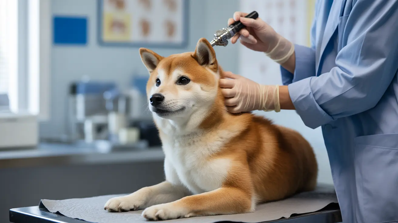
Diagnosis and Treatment Approaches
Veterinarians diagnose posterior synechiae through a comprehensive eye examination using specialized equipment. They'll assess the extent of the adhesions and look for underlying causes that need addressing. Treatment typically involves:
- Medications to dilate the pupil (like atropine)
- Anti-inflammatory drugs
- Treatment of underlying conditions
- Possible surgical intervention in severe cases
- Regular monitoring and follow-up care
Long-term Management and Prevention
Managing posterior synechiae requires a committed approach to eye health. Prevention strategies include regular eye examinations, prompt treatment of eye infections, and protecting your dog's eyes from injury. For dogs with chronic eye conditions, working closely with a veterinary ophthalmologist can help prevent complications.
Frequently Asked Questions
What are the common causes of posterior synechiae in dogs?
The most common causes include chronic eye infections, trauma, severe inflammation (uveitis), corneal ulcers, and surgical complications. Any condition that causes significant inflammation in the eye can potentially lead to posterior synechiae.
How is posterior synechiae in dogs diagnosed, and what are the typical symptoms?
Diagnosis involves a thorough ophthalmic examination using specialized equipment. Common symptoms include squinting, excessive tearing, changes in iris appearance, and visible adhesions. Veterinarians may also use fluorescein dye and measure intraocular pressure during diagnosis.
Can posterior synechiae in dogs lead to glaucoma, and what are the treatment options if it does?
Yes, posterior synechiae can lead to glaucoma by interfering with normal fluid drainage in the eye. If glaucoma develops, treatment options may include pressure-reducing medications, laser surgery, or other surgical interventions to restore proper fluid flow.
How can I prevent synechiae from forming in my dog's eyes?
Prevention focuses on prompt treatment of eye infections, regular veterinary check-ups, protecting eyes from injury, and immediate attention to any signs of eye problems. Early intervention in eye conditions can help prevent synechiae formation.
What are the treatment options for iris bombé (complete posterior synechiae) in dogs?
Iris bombé typically requires surgical intervention to break the complete adhesion. Treatment may include medications to reduce inflammation and pressure, along with surgery to restore normal anatomy and prevent complications like glaucoma.


